 Pathology Training Institute in Bangladesh Best Pathology Training Institute in Bangladesh


Nipah Virus 01797522136
Introduction
Infectious diseases are a major threat to human health in the current world. Especially emerging viral diseases are posing a serious risk to public health. One such dangerous and deadly disease is Nipah Virus Infection. This virus has repeatedly caused outbreaks in various countries of South Asia, including Bangladesh, and it is a matter of special concern as the mortality rate is very high. HRTD Medical Institute, as a renowned medical and diagnostic training institute in Dhaka, is presenting this article with the aim of increasing public awareness and spreading correct medical knowledge. Here, the origin, transmission mode, symptoms, diagnosis, treatment, prevention and its impact on public health of Nipah virus are discussed in detail.

What is Nipah Virus?
Nipah Virus (NiV) is a zoonotic virus, meaning it is transmitted from animals to humans. This virus belongs to the Paramyxoviridae family and belongs to the Henipavirus genus.
Nipah virus mainly causes:
High fever
Difficulty breathing
Encephalitis
Unconsciousness
Can even cause death
The mortality rate of this disease ranges from 40% to 75%, making it one of the most dangerous viruses in the world.
History of Nipah virus
Nipah virus was first identified in Malaysia in 1998–1999. At that time, an unknown encephalitis disease was seen in people associated with pig farms. Later studies revealed that it was a new virus, which was named Nipah virus—after the name of the area called Sungai Nipah in Malaysia.
Later outbreaks of this virus were seen in:
Bangladesh
India (West Bengal and Kerala)
Especially in Bangladesh, Nipah virus has shown a tendency to be transmitted from person to person, which makes it more dangerous.
Nipah virus outbreak and spread
Global situation
Nipah virus is mainly seen in:
Bangladesh
India
Malaysia (in the past)
Singapore (related to the Malaysian outbreak)
Nipah virus in Bangladesh
In Bangladesh, isolated cases of Nipah virus are reported almost every year. The main reasons for this are:
Drinking raw date palm juice
Close coexistence with bats
Lack of awareness in rural areas
HRTD Medical Institute emphasizes on educational training about Nipah virus in Bangladesh.
Natural reservoir of Nipah virus
The main natural reservoir of Nipah virus is:
Fruit-eating bats (Pteropus species)
Although these bats are not sick themselves, they can spread the virus through:
Saliva
Urine
Feces
.
Routes of Nipah virus infection
- Animal to human transmission
Eating bat-eaten fruit
Drinking raw date juice contaminated by bats
Contact with infected animals - Human to human transmission
Being in close proximity to the patient
Coughing or sneezing
Contact with body fluids - Hospital-based transmission
Non-use of PPE
Lack of proper infection control measures
This is why HRTD Medical Institute places great importance on infection control and biosafety training for healthcare workers.
Pathogenesis of Nipah virus
The pathogenesis of Nipah virus infection involves viral entry, widespread dissemination, endothelial damage, and infection of the central nervous system, leading to severe encephalitis and respiratory disease.
1. Entry into the body
- Infection occurs through:
- Contact with infected bats or pigs.
- Consumption of food contaminated with bat secretions (e.g., raw date palm sap).
- Close contact with infected individuals.
- The virus enters through the respiratory tract or oropharyngeal mucosa.
2. Initial replication
- The virus replicates in the epithelial cells of the respiratory tract and nearby lymphoid tissues.
- Early replication allows the virus to spread into the bloodstream.
3. Viremia and systemic spread
- The virus enters the bloodstream (viremia) and disseminates to multiple organs, including:
- Brain
- Lungs
- Spleen
- Kidneys
- Heart
4. Endothelial cell infection
- The virus binds to ephrin-B2 and ephrin-B3 receptors, which are highly expressed on endothelial cells and neurons.
- Infection of endothelial cells causes:
- Vasculitis (inflammation of blood vessels)
- Endothelial injury
- Thrombosis
- Tissue ischemia and necrosis
5. Central nervous system involvement
- The virus crosses the blood–brain barrier and infects neurons.
- This results in:
- Encephalitis
- Neuronal degeneration
- Brain edema
- Microinfarctions due to vasculitis
6. Respiratory disease
- Infection of lung tissue leads to:
- Interstitial pneumonia
- Acute respiratory distress syndrome (ARDS) in severe cases
- Patients with significant respiratory involvement may shed higher amounts of virus, facilitating person-to-person transmission.
7. Immune response
- The innate and adaptive immune systems respond to infection.
- However, the virus can suppress interferon responses, allowing continued viral replication.
- Excessive inflammation may contribute to tissue damage.
Summary flow
Exposure → Entry via respiratory/oral mucosa → Local replication → Viremia → Infection of endothelial cells (vasculitis) → Spread to brain and lungs → Encephalitis + respiratory disease → Severe illness or death
Key pathological features
- Widespread vasculitis involving small blood vessels.
- Endothelial cell infection with thrombosis and necrosis.
- Severe encephalitis with neuronal damage.
- Pulmonary inflammation and edema.
- Multiorgan involvement in severe disease.
After Nipah virus enters the body:
First causes respiratory tract infection
Attacks cells inside the blood vessels
Crosses the blood-brain barrier
Causes inflammation in the brain
Results in:
Acute encephalitis
Respiratory failure
Multiple organ failure
Symptoms of Nipah virus
Incubation period
Usually 4–14 days
Sometimes up to 45 days
Initial symptoms
Fever
Headache
Body aches
Vomiting
Respiratory symptoms
Cough
Shortness of breath
Pulmonary infection
Neurological symptoms
Dizziness
Seizures
Unconsciousness
Coma
Diagnosis of Nipah virus
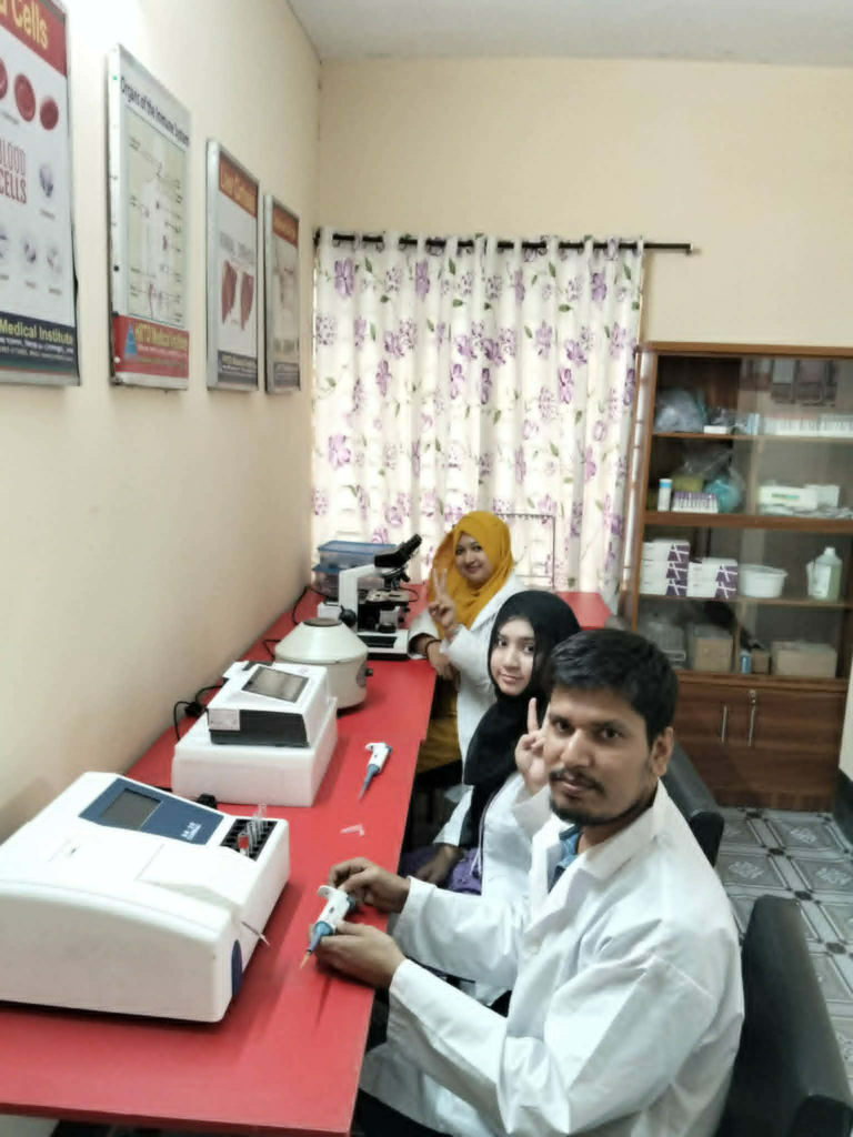
Advanced lab facilities are required for diagnosis of Nipah virus.
Tests
RT-PCR (Most Reliable)
RT-PCR (specifically real-time reverse transcription polymerase chain reaction, or rRT-PCR) is the primary laboratory method used to identify Nipah virus infection by detecting the virus’s RNA in clinical samples.
Here’s how it works:
- Sample collection: Common specimens include throat or nasal swabs, cerebrospinal fluid (CSF), blood, or urine, depending on the stage of illness.
- RNA extraction: Viral RNA is isolated from the sample.
- Reverse transcription: The viral RNA is converted into complementary DNA (cDNA).
- PCR amplification: Specific primers and probes targeting genes of the Nipah virus genome (such as the N, G, or P genes) are used to amplify and detect the viral genetic material.
- Result interpretation:
- Positive RT-PCR: Viral RNA is detected, indicating current infection.
- Negative RT-PCR: Viral RNA is not detected. If testing is done very early or late in the illness, repeat testing or additional methods may be needed if clinical suspicion remains high.
Why RT-PCR is used
- High sensitivity and specificity.
- Can detect infection early, often before antibodies develop.
- Provides relatively rapid results (typically within several hours once the sample reaches a qualified laboratory).
Other diagnostic methods
Additional tests may include:
- Detection of Nipah virus-specific IgM and IgG antibodies by ELISA.
- Virus isolation (performed only in specialized high-containment laboratories).
- Genome sequencing for confirmation or outbreak investigation.
Because Nipah virus is a highly hazardous pathogen, testing is performed only in appropriately equipped reference laboratories with strict biosafety procedures.
ELISA (IgM, IgG)
ELISA (Enzyme-Linked Immunosorbent Assay) is a laboratory test used to detect antibodies produced against Nipah virus. It is commonly used to support the diagnosis of Nipah virus infection, particularly after the immune response has developed.
IgM ELISA
- Detects IgM antibodies, which are the first antibodies produced after infection.
- Usually become detectable within the first week after symptom onset.
- A positive IgM result suggests a recent or current infection.
IgG ELISA
- Detects IgG antibodies, which appear later than IgM.
- IgG antibodies can persist for months or years.
- A positive IgG result indicates past infection, a later stage of infection, or evidence of previous immune exposure.
Interpretation
| IgM | IgG | Interpretation |
|---|---|---|
| Positive | Negative | Early/recent infection |
| Positive | Positive | Current or recent infection with developing immunity |
| Negative | Positive | Past infection or recovery |
| Negative | Negative | No detectable antibodies, or testing performed too early |
Clinical use
- RT-PCR is preferred during the early phase of illness because it detects the virus directly.
- ELISA (IgM and IgG) is especially useful when RT-PCR is negative later in the illness or to confirm infection by demonstrating the body’s antibody response.
- Results should always be interpreted together with the patient’s symptoms, exposure history, and other laboratory findings.
Virus Isolation (High Biosafety Lab)
Virus Isolation (High Biosafety Laboratory)
Virus isolation is the process of growing live Nipah virus from a patient’s clinical specimen. It is considered a definitive method for confirming infection but is performed only in specialized laboratories because the virus is highly infectious.
Principle
Clinical specimens (such as throat or nasal swabs, blood, cerebrospinal fluid, or urine) are inoculated onto susceptible cell cultures. If live virus is present, it infects the cells and replicates, producing characteristic cytopathic effects (CPE). The isolated virus is then confirmed using methods such as RT-PCR, immunofluorescence, or genome sequencing.
Biosafety requirements
- Must be performed in a Biosafety Level 4 (BSL-4) laboratory.
- Requires:
- Maximum biological containment.
- Positive-pressure protective suits.
- Specialized air filtration (HEPA).
- Strict decontamination and waste disposal procedures.
- Highly trained personnel.
Advantages
- Confirms the presence of live infectious virus.
- Enables detailed research on viral characteristics.
- Useful for genome sequencing, antiviral testing, and vaccine development.
Limitations
- Requires BSL-4 facilities, which are limited in number.
- Time-consuming (several days).
- High cost and significant biosafety risks.
- Not routinely used for rapid clinical diagnosis.
Clinical role
For patients with suspected Nipah virus infection, real-time RT-PCR is the preferred diagnostic test because it is faster and safer. Virus isolation is mainly reserved for research, reference laboratories, and outbreak investigations.
Specimen
Blood
CSF
Gallar Swab
Urine
HRTD Medical Institute provides modern diagnostic concepts to Pathology and Lab students.
Treatment
Currently for Nipah virus:
❌ No specific treatment or approved drug
Supportive care
ICU support
Oxygen or ventilation
Seizure control
Prevention of Nipah virus
At the individual level
Do not consume raw date juice
Wash fruits thoroughly
Avoid contact with sick people
In healthcare
Use of PPE
Isolating patients
Hand washing habits
Nipah virus and public health
Nipah virus is a Priority Disease according to WHO, because of:
High mortality rate
Human-to-human transmission
Pandemic potential
Role of HRTD Medical Institute
HRTD Medical Institute:
Produces skilled lab technologists
Provides training in infectious diseases
Leader in pathology and diagnostic education
Address:
Section-6, Block-Kha, Road-1, Plot-11,
Metro Rail Pillar-249, Folpotti Mosque Lane,
Mirpur-10, Dhaka-1216
Contact:
📞 01797522136 | 01987073965 | 01784572173
Nipah Virus Infection
Structure of Henipa Virus
Specialties
Infectious Diseases
Symptoms
Fever, cough, headache
Complications
Encephalitis, seizures
Onset of illness
5-14 days
Cause
Nipah Virus
Diagnosis methods
Symptom-based, confirmed by laboratory tests
Prevention
Stay away from bats and sick pigs, avoid drinking impure date juice
Treatment
Supportive care
Incidence rate
582 people (2001 to 2018)
Conclusion
Nipah virus is a dangerous but preventable disease. With proper knowledge, awareness and trained health workers, the risk of this disease can be reduced to a great extent. HRTD Medical Institute is committed to preparing the future healthcare workforce।